Description
MIKMED-6 variant 74-ST
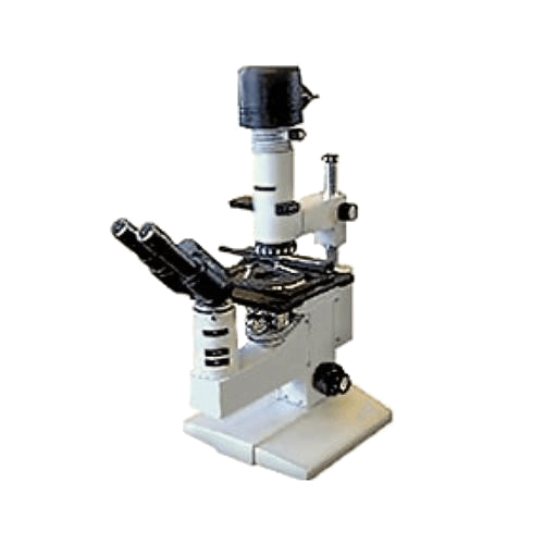
The medical microscope MIKMED-6 version 74-ST is intended for clinical laboratory diagnostics and morphology in the study of objects in transmitted light with illumination using the bright field method, with additional accessories – using phase contrast and dark field methods, in luminescence light and in polarized beams.
In clinical laboratory diagnostics, the microscope MIKMED-6 variant 74-ST is used in blood analysis and viewing cytological preparations of various biological materials: sputum, urine, cerebrospinal fluid, bone marrow, lymph node prints, for the diagnosis of venereal diseases, tuberculosis, in dermatology, as well as for quantitative assessment of the material (leukocyte formula, cytogram, myelogram, scatological analysis, etc.).
On the MICMED-6 microscope, it is possible to study stained and unstained preparations in the form of smears and histological sections, as well as biological fluids in Goryaev-type chambers.
Microscope MIKMED-6 is included in the state register of medical devices, registration certificate FSR 2010/08205, certificate of conformity
ROSS RU.IM32.N00 506 under the scheme of voluntary certification of a medical device.
ADVANTAGES OF MIKMED-6 version 74-ST in comparison with the previous model:
– Well-known manufacturer, mass production at the factory;
– Unified tripod for microscopes of the “Mikmed” series – according to patent No. 84195;
– Objectives of planochromatic correction, tube length – infinity;
– Field of view in image space, increased to 22 mm;
– A five-cavity revolver with a comfortable inclination “from the observer”
– A two-coordinate table with a range of movement of 79×54 mm and the possibility of placing two preparations at the same time, the working surface of the table with a special coating designed for disinfection and resistant to abrasion;
– Illumination system according to Keller, which provides illumination of the fields of view of lenses with a magnification from 4x to 100x without additional adjustments;
– Coaxial mechanism for two-stage coarse and fine focusing;
– Possibility of equipping the microscope with a fast refocusing device;
– Ability to control the coordinate object table with the left hand;
– 12V 20W halogen lamp or 5W LED with 30,000 hours of operation as light source; a secondary power supply with smooth brightness control is built into the microscope stand;
– Possibility of equipping the microscope with a light manager;
– Removable block of the collector assembly, moving forward along the guide slide, providing optimal thermal conditions and convenient safe lamp replacement;
– Compliance with the requirements of the international electrotechnical commission.
By additional order, to expand the possibilities of research, the following options can be included in the microscope:
- Magnifications lenses: 2, 20, 50 mi, 60;
2. Eyepieces with diopter adjustment mechanism: 10/22 with scale, 15/16, 30/8;
3. Device for observation by the method of phase contrast and dark field;
4. Dark field condenser;
5. Simple polarization device;
6. Attachment with an illuminator for studying objects in the light of luminescence, including one based on a monochromatic LED instead of a mercury lamp;
7. High resolution digital camera with adapter (C-mount) for visualization and computer analysis;
8. Fast refocusing device;
9. Transferring the coaxial handles of the table to control the left hand.
Specifications
| NameOption of configuration MIKMED-6-74-ST | Specifications |
| Apparent magnification, times | from 40 to 1000 |
| Lenses – tube infinity, magnification / aperture | planachromatic correction4x/0.10; 10x/0.25; 40x/0.65; 100x/1.25 mi |
| Wide-field eyepieces, visible magnification, multiple/field, mm | with a diopter mechanism 10x/22, the ability to work with glasses |
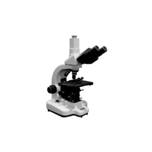
| Observation Attachment | trinocular |
| Binocular tube Siedentopf | does not require adjusting the length of the tubes when installing the eye base |
| Tilt of eyepiece tubes | 30 deg. |
| Interpupillary distance | adjustable from 48 to 75 mm |
| Changing the working height of the microscope (distance from the base of the microscope to the exit pupil) | by adjusting the position of the exit pupils by means of eyepiece tubes |
| Parallelism of the beams emerging from the nozzle eyepieces in the directions: | vertical – divergence no more than 15′, horizontal – convergence no more than 20′, – divergence no more than 60′ |
| Revolver 5-cavity, rotation in any direction | for the convenience of changing preparations, it is inclined to the tripod from the “observer” |
| 2-coordinate object stage, movement range |
equipped with a glass-ceramic insert coaxial handles for the right hand, 78×54 mm, the installation of two preparations at the same time |
| Bright field condenser with iris aperture diaphragm | numerical aperture 0.9/1.25 mi |
| Lighting – firmware (Koehler), | centerable and focusable iris field diaphragm |
| Manifold assembly with vents, removable, slides forward on guide slides | provides: optimal thermal conditions; convenient and safe replacement of the light source |
| Light source white LED | 5 W run time 30,000 hours |
| The power supply is built into the tripod, provides smooth brightness control | complies with the established standards: – electrical safety (class I, type H according to GOST 12.2.091, GOST R 51350 (IEC 61010-1), – electromagnetic compatibility (GOST R IEC 61326-1), – radio interference (GOST R 51318.15.) |
| Light manager for operational control of the level of illumination in the plane of the object. | 5-level light manager system, manager LEDs located on both sides of the base |
| Power consumption, VA, no more | 25 |
| Overall dimensions of the microscope, mm, no more | 270x380x450 |
| Weight, kg, no more | 8.5 |
| Overall dimensions of the microscope, in the package, mm, no more | 300x400x530 |
| Packed weight, kg, no more | 10 |
| Certificate of conformity | according to the scheme of voluntary certification |