Description
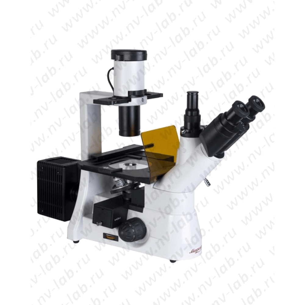
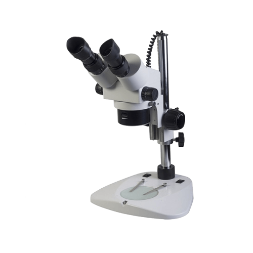
Trinocular inverted biological microscope Mikromed I – is designed for studies of low-contrast cell cultures of tissues, sediments of liquids, etc., located in a special container.
Inverted trinocular microscope Mikromed I is designed to study objects in transmitted light by the bright field method, as well as by the phase-contrast method.
Distinctive features:
- The thickness of the object of study does not play a role, since the inverted design of the microscope p (illumination of the object from above, observation – from below) allows you to examine dimensional objects or objects located in special dishes (Petri dishes, flasks, etc.), as well as viewing the nutrient medium under the monolayer.
- High-contrast optics for flat object imaging and comfortable microscope work
- It is possible to install visualization systems: video eyepieces, a visualization kit based on a Canon A 640 digital camera
Specifications:
- Visible magnification of the microscope, multiple: 40-400
- Visible magnification of wide-field eyepieces, multiple: 10
- Plan lens magnification: 4,10,20,40
- Lens Working Distance: 1.5mm
- Revolving device: “reverse” – revolver for 5 lenses
- Adjustable interpupillary distance, within: 50-80 mm
- The angle of inclination of the eyepiece tubes of the nozzle, degrees: 45
- Numerical aperture of the condenser: 0.6
- Dimensions of the subject table, mm: 170×240
- Light source: Halogen lamp 12V 30W
- Microscope power: AC 220 V; 50/60 Hz
Advantages:
- Ergonomic tripod design with coaxial coarse and fine focus handles
- Improved image quality and comfort of observation of the object under study due to the use of high-quality optics that provide a flat field of view of the microscope, which is increased to 22 mm.
- Ability to use visualization systems
Areas of use:
- Medicine
- Various areas of biology; biotechnology
- pharmaceutical industry
- Virology
- Hydrobiology
- Agriculture
- Ecology
| Name | Quantity | Note |
| Microscope stand with built-in power supply and trinocular attachment | 1 | Trinocular head magnification – 1 |
| Illuminator | 1 | |
| Carriage with 2 framed light rings and window for transmitted light | 1 | Light rings are designed for observations: by the phase contrast method with phase-contrast objectives 10x, 20x |
| Lens 4x/0.10 ∞/- Plan | 1 | |
| Lens 10x/0.25 ∞/- Plan | 1 | phase |
| Lens 20x/0.40 ∞/1.5
L Plan |
1 | phase |
| Lens 40x/0.60 ∞/1.5
L Plan |
1 | |
| Wide-field eyepieces x/field, mm: 10x/22 | 2 | To work with glasses |
| Inserts on the subject table | 2 | one glass |
| Light filters | 4 | Blue; green; yellow, matt |
| Eyecup for eyepiece | 2 | |
| Microscope “ST” | 1 | |
| Halogen lamp | 2 | 12V 30W |
| Fuse (fusible insert) | 2 | 1 A |
| Manual | 1 | |
| Usability
visualization systems |
Eat | Provided by the presence of a vertical tube – visualization channel |

