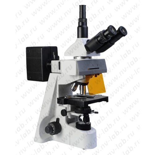
Description
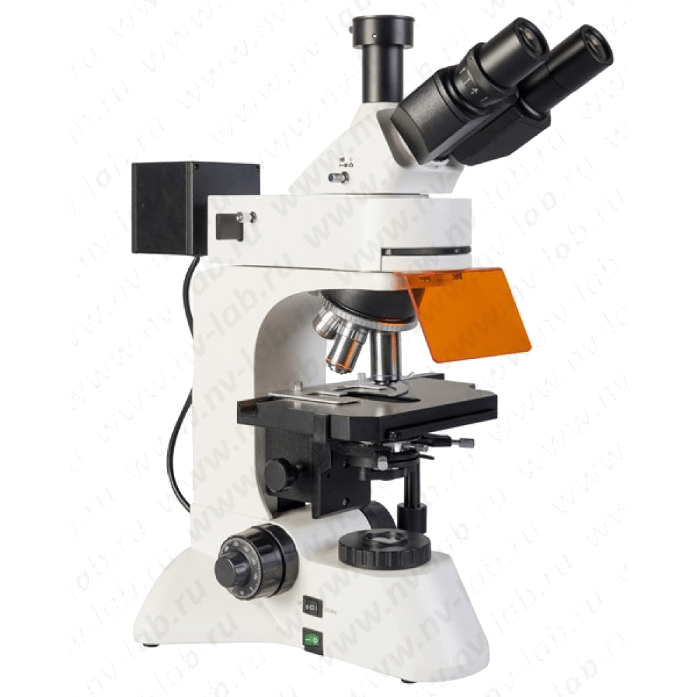
Micromed 3 LUM LED
Mikromed 3 LUM LED – LED luminescent microscope is easy to use, provides fast, highly sensitive research.
The trinocular luminescent microscope MICROMED 3 LUME LED is designed to observe the image of objects in the light of visible luminescence, as well as in transmitted light in a bright field. In addition to the possibility of studying objects in transmitted light (classical illumination according to Keller), the following types of studies are carried out using fluorescence techniques on a microscope MICROMED 3 LUM LED: immunochemical, immunological, immunomorphological, immunogenetic.
In the process of researching drugs – blood smears, bone marrow, tissue sections, hidden infections are detected, such as chlamydia, ureaplasmosis, mycaplasmosis, herpes and others; as well as the detection of differentiation antigens T and B of lymphocytes, atypical blood cells; express diagnostics of bacterial, viral, protozoal, etc. infections; determination of antinuclear factor and the like; immunochemical diagnosis of leukemia; chromosome analysis.
The microscope is also used in veterinary medicine, crop production, biotechnology, the pharmaceutical industry, for examinations in the field of forensic science, sanitary and epidemiological surveillance, and environmental protection.
On the microscope, images of objects can be photographed using a F/C-based imaging kit (not included) and the image can be displayed in real time on a PC screen using a video eyepiece (not included).
The main advantages of the microscope Mikromed 3 LUM LED
- The kit includes 4 luminescent blocks (UV, V, B, G)
- The source of both transmitted and reflected light are LEDs – cost-effective energy consumption and ease of use.
- Quick switching of research modes – from the luminescence method to the bright field method.
- Modern ergonomic design.
- The optical scheme of the microscope is designed for infinity
- The eyepieces have a field of view of 22 mm, diopter correction of vision and a “remote pupil”, which makes it equally convenient to work both with and without glasses.
- Planachromat lenses provide a flat image of an object across this field of view
- Base with built-in power supply for transmitted light illuminator and fluorescent illuminator
- High-precision assembly and adjustment of the microscope, parfocal objectives
- The revolving device is turned away from the observer
- The spring frame for lenses with a magnification of 40x and 100x provides protection from mechanical damage to the object and the front lens of the lens
- 2D stage with coaxial handles
- Coaxial coarse and fine focus mechanism
- Coarse focus locking mechanism for quick microscope adjustment when changing specimens. Coarse focus hardness adjustment
Characteristics
| Microscope magnification, times | 40 – 1000 |
| Spectral range of luminescence excitation, nm | 330 – 550 |
| Spectral range of the studied luminescence, nm | 425 – 700 |
| visual nozzle | trinocular |
| Angle of inclination of the visual nozzle, hail | thirty |
| Adjustable interpupillary distance, within, mm | 55-75 |
| Nozzle enlargement | 1 |
| Eyepieces | wide-field with remote pupil 10/22 |
| Revolving device | for 5 lenses |
| Lens Correction Type | Planachromats, for work in the light of visible luminescence, designed for the length of the tube “infinity” |
| Lenses | 4x/0.1; 10x/0.25; 40x/0.65; 100x/1.25 mi |
| Subject table, mm | 210×140 |
| Range of drug movement, mm | 75×50 |
| Abbe centered condenser, max. numerical aperture | 1.25 |
| transmitted light source | high power dimmable LED |
| Fluorescent Light Source | high power dimmable LED (4pcs) |
| Power supply – AC, V / Hz | 220+-22/50 |
| Overall dimensions, mm | 520x460x250 |
| Weight, no more than, kg | 14 |

