Description
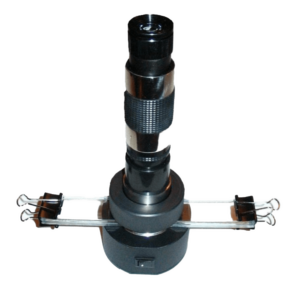
The portable trichinelloscope PT-101 is designed for diagnosing trichinosis in wild and domestic animals by compression trichinoscopy.
Trichinelloskop can be used in laboratories of veterinary sanitary examination, veterinary institutions, farms and hunting farms, as well as private producers of meat products.
The device makes it possible to detect parasites in fish muscles, trematode partenids in freshwater mollusks during helminthological assessment of pastures, and can also be used as a conventional microscope. All observations are made in transmitted light.
Technical characteristics of trichinelloscope PT-101:
- Magnification – 50x times.
- Field of view diameter 8 mm.
- Operating time from one battery is not less than 60 hours.
- Overall dimensions of the trichinelloscope device in the working position without installed slides (no more than) diameter 73 mm, height 240 mm.
- The mass of the device is not more than 0.5 kg.
- The mass of the device in the package is not more than 0.7 kg.
- Power consumption 0.09 Watts.
- Average service life is 3 years.
- Mean time between failures is not less than 5000 hours.
- The operating temperature of the trichinelloscope is from -20°C to +50°C at a relative humidity of not more than 80%.
- The trichinelloscope is transported in a packing bag.
- The warranty period of the portable trichinelloscope is 1 year from the date of shipment from the manufacturer.
Contents of delivery:
- Trichinelloscope PT-101
- Microscope slide – 2 pcs.
- Cover glass compressor – 2 pcs.
- “Krone” type battery – 1 pc. Photograph of Trichinella larvae in pig meat
- Clip – 4 pcs.
- User manual – 1 pc.
- Medical gloves – 1 pair.
- Packing bag – 1 pc.
- Medical scissors – 1 pc.
The procedure for determining trichinosis:
- From each muscle sample of the tested animal, cuts are made strictly along the muscle fibers with a size of 5×2 mm. In total, 24 cuts are made from different parts of the sample, and with negative results, 3-4 times more.
- Each slice is placed on a separate sector of the compression slide (a total of 8 sectors on the slide).
- The compressor is covered with the top glass.
- Squeeze and remove excess liquid to prevent it from getting inside the device.
- The glasses are placed in the trichinoscope and fixed from different sides with clamps.
- The slides are firmly pressed against the base of the trichinelloscope using a clamping ring. In the absence of compressor, it is possible to use glass with a thickness of 3-4 mm and a width of not more than 25 mm.
- Each of the prepared samples is microscoped separately. Sharpness is set by rotating the eyepiece and objective.
Microscopy result:
the encapsulated larva of Trichinella in the muscles is a capsule of the larva of a lemon-shaped or oval shape 0.5-0.7 x 0.2-0.3 mm in size; inside the capsule one, rarely two or three spirally folded larvae. With the non-capsular variant, only spirally folded Trichinella larvae are found.
If at least one Trichinella larvae is found in the muscles, all muscle samples are disposed of, before that they fall asleep or pour disinfectants – bleach or bleach! Compressor glasses are disinfected by boiling for 60 minutes from the moment of boiling.


